
PACS and RIS: The Nervous System of Modern Radiology
PACS and RIS: the nervous system of modern radiology
In the previous article, “How medical imaging systems have evolved”, we explored the fascinating journey from the first X-rays to the digital and intelligent era of medical imaging. But that technological revolution would not have been possible without the systems that give life, order and coherence to that entire flow of information: PACS and RIS, the true nervous systems of modern radiology.
From film to data: the birth of PACS
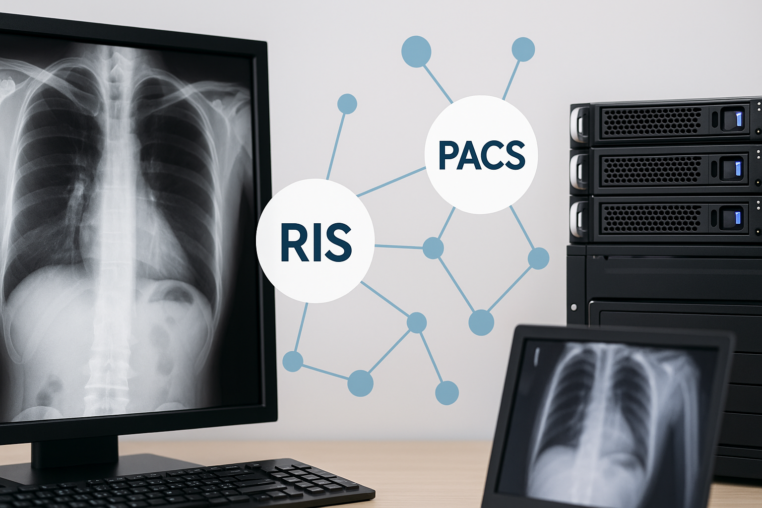
In the analog era, hospitals were full of yellow envelopes stuffed with physical X-rays. Every image had to be developed, filed and carried by hand. It was a slow, costly and vulnerable process. Everything changed with the arrival of PACS (Picture Archiving and Communication System), a technology that made it possible to store, view and share medical images in digital format.
PACS not only did away with cabinets full of films, it also connected radiology with the entire hospital. Physicians, surgeons and specialists can access images from anywhere, at any time, and make decisions in real time. It is the bridge between technology and medical care.
RIS: the organizing brain
If PACS is the nervous system of radiology, the RIS (Radiology Information System) is its brain. This system manages the administrative and logistical side of the process: study scheduling, patient registration, reports, follow-up and communication between departments.
The RIS coordinates the workflow, assigns priorities, links images to the patient's medical record and integrates with other hospital systems such as the HIS (Hospital Information System). Without RIS, modern radiology would be a set of uncoordinated pieces.
When PACS and RIS work together
The real power emerges when both systems are integrated. While the RIS manages patient information and medical reports, the PACS manages the associated images. Together, they create a complete digital ecosystem where every study has a clinical context, every image a diagnosis and every decision a solid data foundation.
This combination enables:
- Immediate access to images and reports from any authorized device.
- Shorter turnaround times for diagnosis and delivery of results.
- Interoperability with other systems and international standards (such as DICOM and HL7).
- Security and traceability in the handling of sensitive data.
PACS and RIS in the age of artificial intelligence
Today, PACS and RIS systems are no longer just storage or management tools; they have become intelligent platforms. They integrate artificial intelligence that analyzes patterns in images, prioritizes urgent studies and helps radiologists detect anomalies in seconds.
This combination of automation and predictive analytics is changing the specialist's role. The radiologist no longer just interprets images: they supervise, validate and complement the findings proposed by AI. It is a collaborative effort between human expertise and algorithmic precision.
The new ecosystem of digital radiology
Modern hospitals are built on this invisible foundation. PACS and RIS cannot be seen, but they connect everything: from the acquisition of an image in an MRI room to the final report in the patient's electronic health record. They are the connective tissue that keeps the technological heart of visual medicine beating.
Thanks to them, radiology is no longer an isolated process and has become a nerve center of medical information that feeds clinical decisions, research and personalized treatments.
Conclusion
PACS and RIS systems are much more than software: they are the backbone of contemporary radiology. They have allowed images to travel at the speed of data and diagnoses to become faster, more accurate and more collaborative. If X-rays were the beginning of medical vision, PACS and RIS are the nervous system that keeps that vision alive in the 21st century.
← Previous: How medical imaging systems have evolved
Next: Automatic tumor detection: algorithms that see the invisible →
Related Articles
Keep exploring similar content.
Augmented Reality and 3D in Surgical Procedures
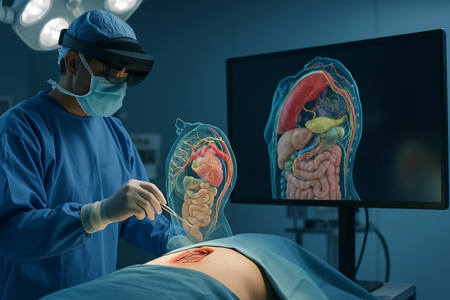
Augmented reality and 3D modeling are transforming how surgical procedures are planned, performed and taught, giving surgeons a digital window into the patient's anatomy.
Data Security in Digital Radiology
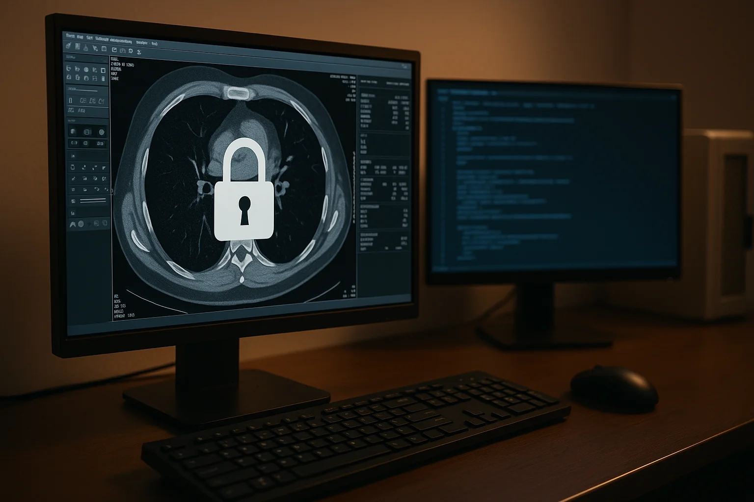
Digital radiology handles sensitive images, predictive models and valuable clinical data. Protecting this information is essential to ensure safe diagnoses, patient trust and operational continuity.
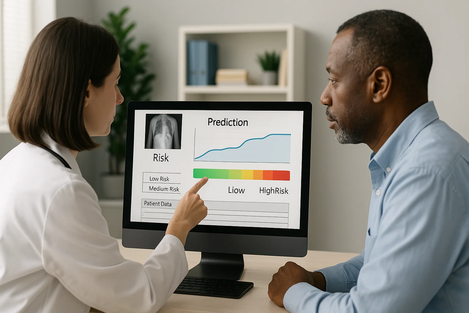
Image-Based Predictions: Medicine That Stays One Step Ahead
Thanks to artificial intelligence, medical images no longer just show what is happening, but what could happen. Predictive medicine turns every study into a tool to anticipate risks and save lives ahead of time.