
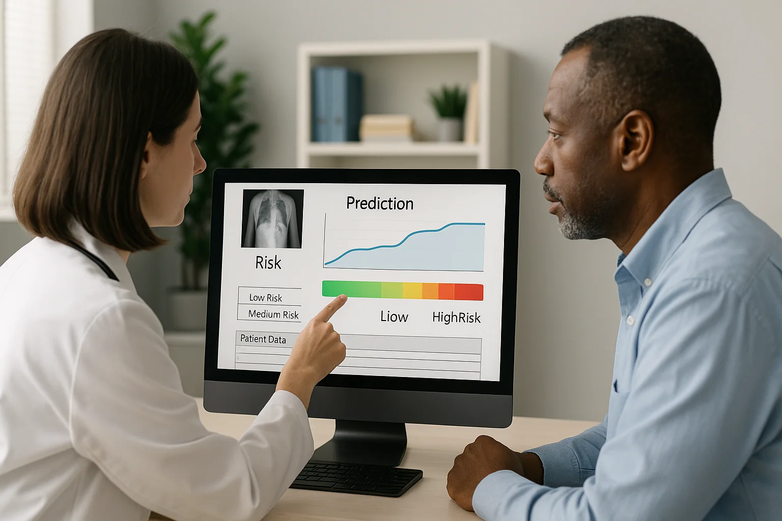
Image-Based Predictions: Medicine That Stays One Step Ahead
Image-based predictions: medicine that stays one step ahead
In the previous article, “Fusing imaging + clinical data: integrated diagnosis” , we analyzed how combining medical images, genetic data, habits and clinical history is building a complete picture of the patient. But when that integration is combined with artificial intelligence, something even more powerful happens: images stop describing the present and begin to predict the future.
It is the birth of a new discipline: image-based predictive medicine.
From seeing the obvious to anticipating the invisible
Medical images —CT, MRI, PET, ultrasound— have been revealing visible lesions for decades. But modern algorithms can detect patterns, textures and micro-signals that do not appear in traditional human interpretation. These patterns, known as radiomics, allow an image to contain information with predictive value.
For example:
- An algorithm can detect the probability that a tumor will grow aggressively.
- It can anticipate the risk of a bone fracture before it happens.
- It can predict future heart failure by analyzing the subtle shape of the ventricle.
We are no longer talking about diagnosis: we are talking about anticipation.
How image-based predictions are created
AI prediction models follow a complex but fascinating process:
- 1. Feature extraction (radiomics): the algorithm converts each pixel or voxel into measurable data.
- 2. Integration of clinical information: genetics, lab results, medical history, habits.
- 3. Training with millions of cases: the AI learns which patterns are associated with future diseases.
- 4. Generation of predictive models: the system calculates risks, trends and the probability of progression.
It is like teaching a machine to see the future based on the past of thousands of patients.
Real examples where prediction is already a reality
Image-based predictive medicine is already saving lives in multiple areas:
- Oncology: prediction of metastasis, response to treatment and survival.
- Cardiology: risk of heart failure, atrial fibrillation, coronary events.
- Neurology: progression of Alzheimer's or mild cognitive impairment.
- Orthopedics: prediction of fracture risk due to hidden bone density loss.
- Pulmonology: progression of lung nodules before they grow.
What used to be discovered too late can now be anticipated months —or even years— in advance.
The key role of integrated diagnosis
As we saw in the previous article, integrated diagnosis combines imaging + clinical data. That integration is essential for prediction. AI does not predict based only on what it sees, but on everything it knows about the patient.
Prediction is not magic: it is clinical mathematics with human context.
Explainable AI: the indispensable bridge
When an algorithm predicts a risk, the physician needs to know why. That is why explainable AI is key to making prediction reliable and ethical.
The new systems show:
- Which areas of the image influenced the prediction.
- Which radiomic features were relevant.
- How clinical data influences the final result.
Prediction is no longer a black box: it is a transparent analysis.
The impact on clinical practice
- Better-informed physicians: they make decisions with evidence about the future.
- Safer patients: they can act before the disease progresses.
- A more sustainable system: the cost of late treatments is reduced.
- Personalized prevention: each patient receives recommendations tailored to their risk.
Prediction turns every imaging study into an opportunity to get ahead of disease.
Conclusion
Medicine that stays one step ahead is no longer science fiction. Thanks to artificial intelligence and in-depth image analysis, healthcare is entering an era in which the future can be calculated, prevented and transformed before symptoms appear. Every pixel becomes a data point, and every data point a clue that makes it possible to save lives in advance.
← Previous: Fusing imaging + clinical data: integrated diagnosis
Related Articles
Keep exploring similar content.
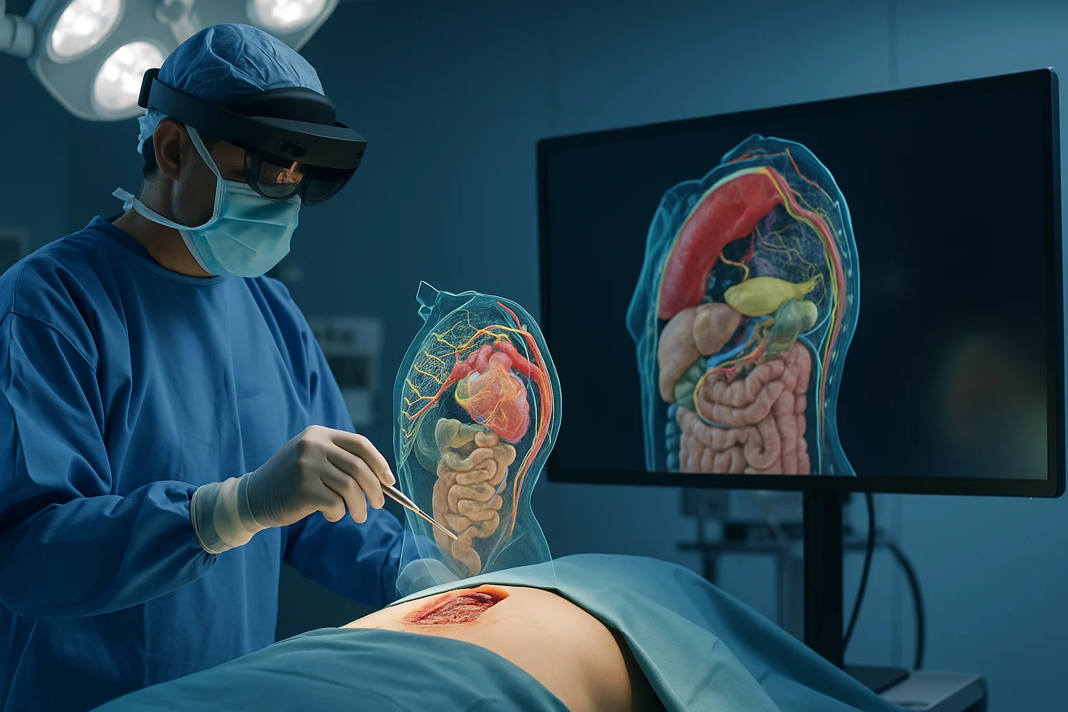
Augmented Reality and 3D in Surgical Procedures
Augmented reality and 3D modeling are transforming how surgical procedures are planned, performed and taught, giving surgeons a digital window into the patient's anatomy.
Data Security in Digital Radiology
Digital radiology handles sensitive images, predictive models and valuable clinical data. Protecting this information is essential to ensure safe diagnoses, patient trust and operational continuity.
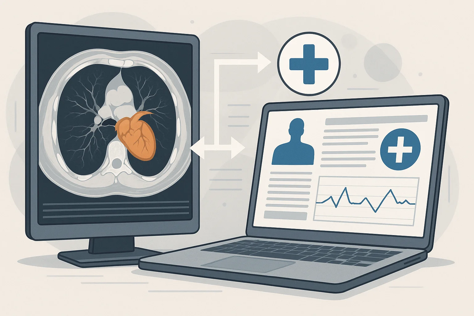
Fusing Imaging and Clinical Data: Integrated Diagnosis
Combining medical images with clinical, genomic and lifestyle data produces more complete and personalized diagnoses, turning precision medicine into a reality.