
Augmented Reality and 3D in Surgical Procedures
Augmented reality and 3D in surgical procedures: when precision outperforms the scalpel
Surgery has always been an art that combines skill, experience and nerves of steel. But in recent years, that art has found a quiet yet powerful ally: augmented reality (AR) and 3D models. These technologies are transforming the way surgical procedures are planned, performed and evaluated around the world.
Today, a surgeon no longer relies solely on clinical judgment: they have a digital window that lets them see internal structures, simulate operations and navigate the human body like never before. Welcome to the new era of augmented surgery.
From imagination to 3D modeling
In the past, a surgeon had to interpret two-dimensional images —such as CT or MRI scans— and mentally reconstruct the patient's anatomy. Now, thanks to 3D modeling, that reconstruction becomes tangible.
Digital models generated from medical images make it possible to:
- Plan surgery step by step.
- Simulate scenarios based on different approaches.
- Study complex cases anatomically with a precision impossible to achieve on paper.
It is like having an exact replica of the patient before picking up the scalpel.
Augmented reality: seeing beyond the visible
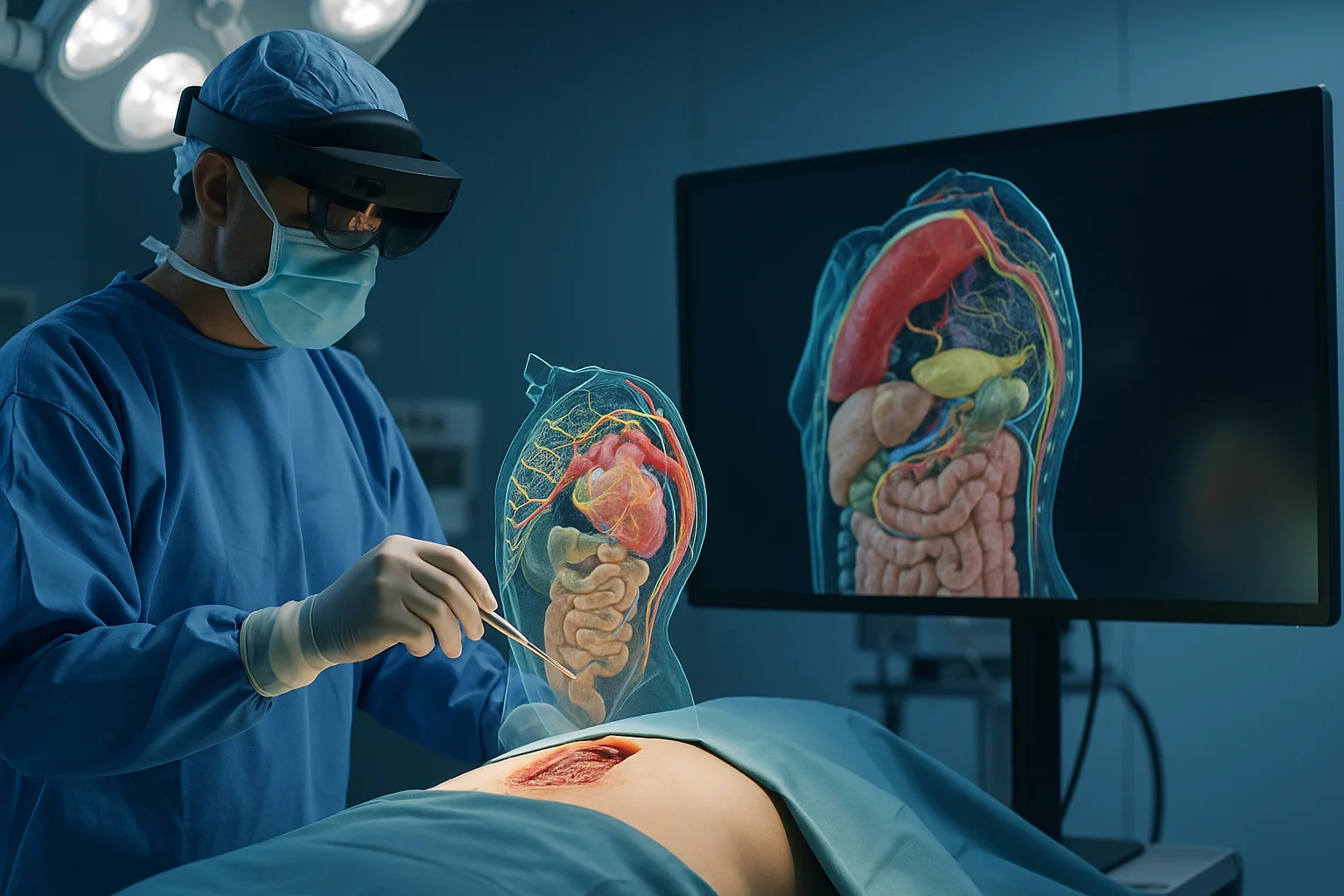
Augmented reality makes it possible to overlay digital information onto the real surgical field. Through special glasses, screens or head-mounted displays, the surgeon can visualize:
- Hidden structures
- Ideal paths for incisions
- Organs, vessels and nerves in real time
- 3D planes superimposed on the real anatomy
It is no longer just about seeing: it is about anticipating.
How it works in practice
The process combines three pillars:
- Medical images (CT, MRI) that provide the anatomical basis.
- 3D reconstruction software that models the anatomy.
- AR devices that project the information in real time.
The surgeon views the surgical area with layers of information that show exactly where to cut, what to avoid and what the safest path is. It is like having a surgical GPS inside the operating room.
Real applications already changing medicine
- Neurosurgery: mapping of tumors and trajectories to avoid critical areas of the brain.
- Orthopedic surgery: placement of prostheses with millimeter precision.
- Oncology: exact localization of tumors invisible to the naked eye.
- Cardiology: navigation within complex vascular structures.
The results: fewer complications, better surgical times and faster recoveries.
AR as a training tool
Residents no longer learn only from books or mannequins: they can practice with digital models that replicate real cases.
- Complete virtual surgeries.
- 3D simulators with haptic feedback.
- Interactive assessments.
Surgical training enters a new dimension in which mistakes do not cost lives and learning is limitless.
AI as a silent co-star
Although this article is about AR and 3D, we cannot forget that artificial intelligence is behind many of these improvements. AI helps outline organs, detect complex structures and generate hyper-precise models. Without it, augmented surgery would be slower, less precise and less accessible.
Adoption challenges
Like any emerging technology, AR and 3D surgery face challenges:
- Equipment costs.
- Learning curves.
- Integration with hospital systems.
- Ongoing clinical validation.
But the trend is clear: every year it becomes more accessible, more intuitive and more precise.
Conclusion
Augmented reality and 3D modeling represent one of the most transformative advances in modern surgery. They make it possible to see the invisible, plan precisely and operate with a level of safety that was previously unthinkable. The medicine of the future no longer depends solely on human skill, but on a perfect alliance between technology, advanced visualization and clinical intelligence.
← Previous: Data security in digital radiology
Next: →
Related Articles
Keep exploring similar content.
Data Security in Digital Radiology
Digital radiology handles sensitive images, predictive models and valuable clinical data. Protecting this information is essential to ensure safe diagnoses, patient trust and operational continuity.
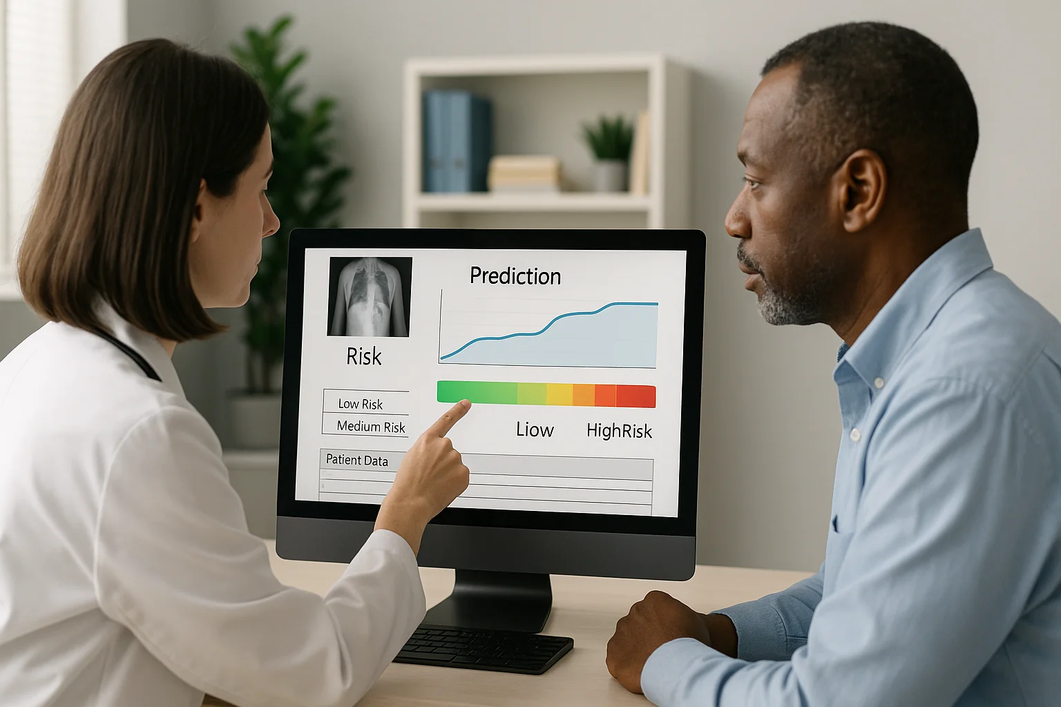
Image-Based Predictions: Medicine That Stays One Step Ahead
Thanks to artificial intelligence, medical images no longer just show what is happening, but what could happen. Predictive medicine turns every study into a tool to anticipate risks and save lives ahead of time.
Fusing Imaging and Clinical Data: Integrated Diagnosis
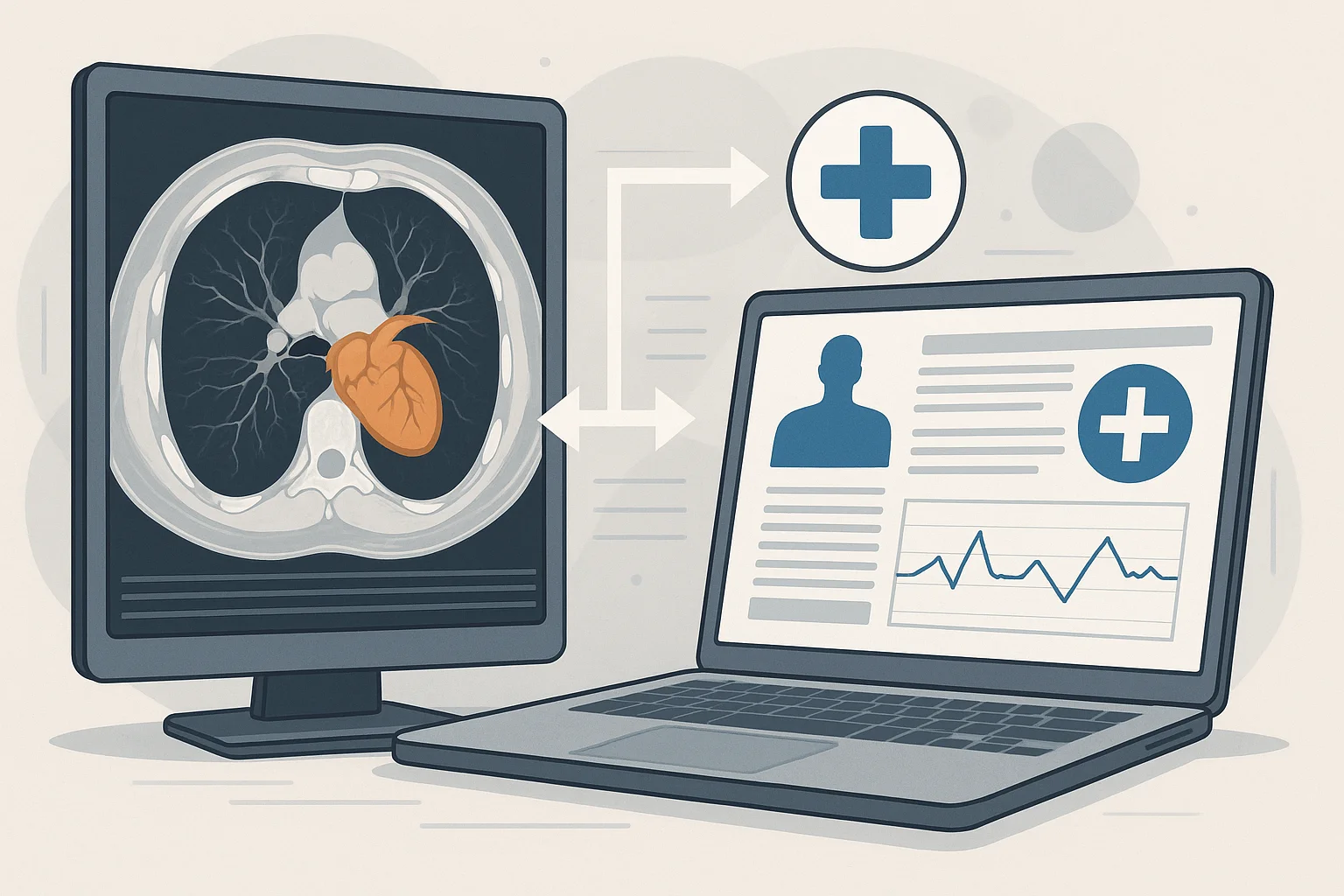
Combining medical images with clinical, genomic and lifestyle data produces more complete and personalized diagnoses, turning precision medicine into a reality.