
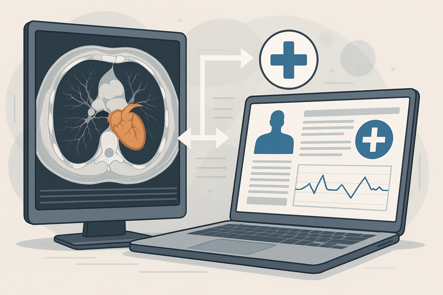
Fusing Imaging and Clinical Data: Integrated Diagnosis
Fusing imaging + clinical data: integrated diagnosis
In the previous article, “How deep learning improves MRI and CT scans” , we saw how deep neural networks not only interpret images but also reconstruct them, correct noise and speed up studies such as MRI and CT scans. That technological leap leads us to an even greater evolution: integrating those optimized images with clinical, genetic and contextual data. It is the birth of integrated diagnosis.
From the organ to the whole organism
Traditionally, radiology analyzed what the image showed: a tumor, a lesion, an anomaly. But the human body does not work in isolated compartments. Today, medicine knows that an image is only one piece of the puzzle. True diagnosis comes from combining the image with everything that is known about the patient.
And that is where the revolution begins: see + understand + contextualize.
What fusing imaging with clinical data means
This integration combines multiple sources of information to obtain a 360° view of the patient:
- Medical images: CT, MRI, PET, ultrasound.
- Electronic health record: medical history, medication, allergies.
- Lab data: tumor markers, hormone levels, blood count.
- Genetic information: mutations, susceptibility to diseases.
- Lifestyle data: sleep, nutrition, physical activity.
- Environmental factors: pollution, climate, occupational hazards.
Each of these data points adds an additional layer of understanding. Diagnosis stops being a snapshot and becomes a complete story.
AI as the architect of diagnosis
The big question is: how do you bring so much information together? The answer lies in machine learning and deep learning algorithms, capable of identifying invisible correlations between clinical data and radiological findings.
For example, a system can:
- Detect a mass on an MRI.
- Cross-reference it with suspicious biomarkers.
- Relate it to previously identified genetic mutations.
- Compare its progression with earlier studies.
- Factor in the patient's lifestyle to adjust their risk.
The end result is not just an interpreted image, but a contextualized diagnosis with personalized recommendations.
Deep learning as a bridge to integration
As we saw in the previous article, deep learning already improves images before the radiologist sees them. But that optimization is not an end in itself: it is the gateway to combining cleaner, more accurate images with high-quality clinical data.
When an image is clear, consistent and standardized, the algorithm can:
- Relate it to global databases.
- Compare it with similar cases.
- Extract features (radiomics) useful for predictive models.
In this way, the image becomes one more data point within the clinical ecosystem.
The radiologist as a knowledge integrator
In this new model, the radiologist no longer just describes findings: they become a multidimensional clinical analyst.
Their role includes:
- Interpreting images together with clinical and genomic data.
- Validating AI suggestions.
- Collaborating in multidisciplinary teams.
- Offering personalized recommendations based on all available evidence.
The image is no longer the end of the diagnosis, but the starting point for an integrated view.
Real cases where fusion is changing medicine
- Precision oncology: tumors analyzed with radiomics + genetic mutations.
- Cardiology: coronary CT + biomarkers + lifestyle habits.
- Preventive medicine: images + longitudinal data + predictive machine learning.
In each case, integration improves accuracy and speeds up critical decisions.
A more human diagnosis thanks to data
Paradoxically, the more we use artificial intelligence, the more human medicine becomes. Integrated diagnosis is not just about data: it is about understanding a person's complete story. The patient stops being “an imaging study” and becomes a unique combination of biology, habits, genetics and environment.
Conclusion
The fusion of imaging + clinical data represents the natural evolution of modern radiology. Deep learning has already improved images; now the challenge is to integrate them into a larger, smarter and more personalized clinical system. The diagnosis of the future will be neither purely visual nor purely statistical: it will be integrated, predictive and deeply human.
← Previous: How deep learning improves MRI and CT scans
Next: Image-based predictions: medicine that stays one step ahead →
Related Articles
Keep exploring similar content.
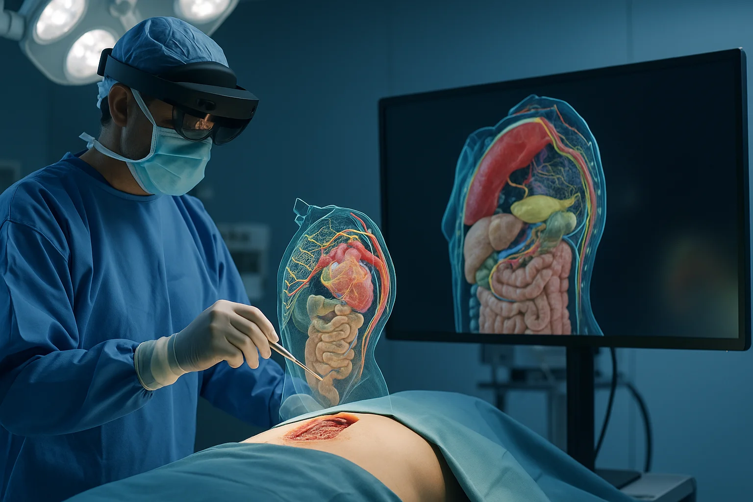
Augmented Reality and 3D in Surgical Procedures
Augmented reality and 3D modeling are transforming how surgical procedures are planned, performed and taught, giving surgeons a digital window into the patient's anatomy.
Data Security in Digital Radiology
Digital radiology handles sensitive images, predictive models and valuable clinical data. Protecting this information is essential to ensure safe diagnoses, patient trust and operational continuity.
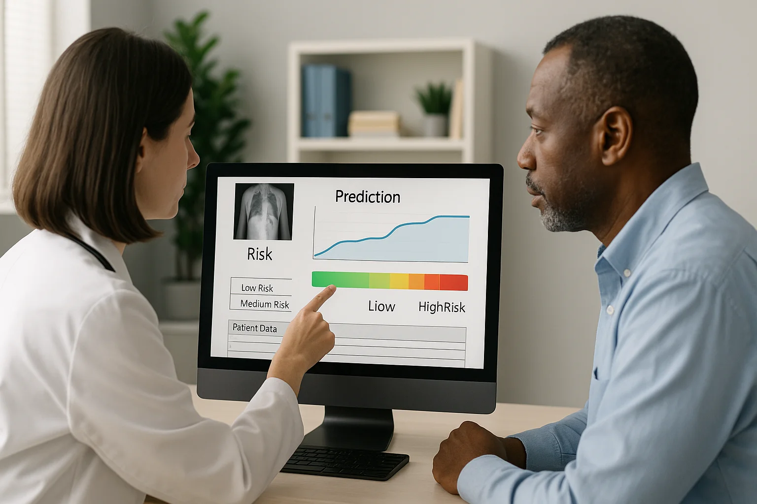
Image-Based Predictions: Medicine That Stays One Step Ahead
Thanks to artificial intelligence, medical images no longer just show what is happening, but what could happen. Predictive medicine turns every study into a tool to anticipate risks and save lives ahead of time.