
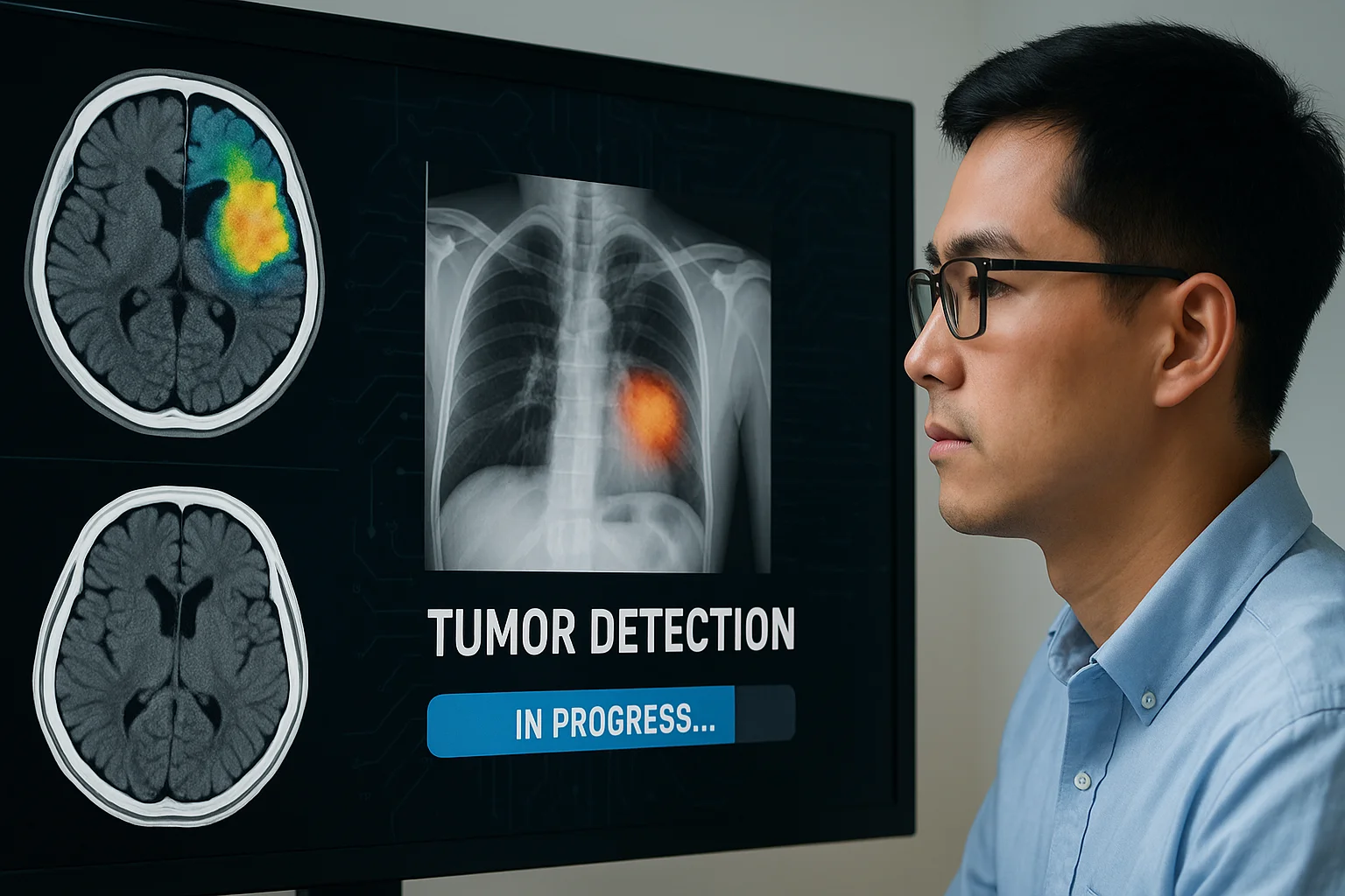
Automatic Tumor Detection: Algorithms That See the Invisible
Automatic tumor detection: algorithms that see the invisible
In the previous article, “PACS and RIS: the nervous system of modern radiology”, we explored how digitization allowed medical images to travel, be shared and be analyzed in real time. Now the revolution goes one step further: algorithms no longer just transport images, they learn to see them.
Automatic tumor detection represents one of the most impressive frontiers of artificial intelligence applied to medicine. We are facing a paradigm shift in which the machine does not replace the physician but empowers them, seeing what the human eye cannot.
When the machine learns to look
Modern algorithms use deep learning techniques and convolutional neural networks (CNNs) to analyze millions of medical images. These networks mimic the structure of the human brain and can learn, on their own, to distinguish between healthy and abnormal tissue. With each new image, their accuracy increases.
The result is striking: current systems can detect microtumors in X-rays, MRIs or CT scans long before they are perceptible to the human eye. And unlike humans, who are subject to tiredness or subjectivity, algorithms never get distracted or fatigued.
How these algorithms work
The process begins with massive training. Thousands or millions of labeled medical images —in which human specialists mark the areas with tumors— are fed into the system. From there, the algorithm “learns” patterns: shapes, textures, densities, colors and contrasts associated with lesions. Over time, it becomes capable of recognizing those signs in new images without human intervention.
The key lies in combining data: the image alone is not enough. The algorithm also cross-references information on age, medical history, genetics and lab results, achieving a contextualized and personalized diagnosis.
Advantages over traditional diagnosis
- Early detection: systems can identify microscopic tumors before they are clinically visible.
- Fewer errors: they minimize variability between human observers.
- Faster diagnosis: they make it possible to process large volumes of images in seconds.
- Support for physicians: they generate visual maps, statistics and automated reports that make decision-making easier.
Real examples of AI in action
Today, hospitals in Europe, Asia and the Americas already use FDA- or CE-certified algorithms to assist in detecting breast, lung, skin or brain cancer. Systems such as Google DeepMind Health, IBM Watson Imaging and Siemens AI-Rad Companion have become allies of the modern radiologist, raising accuracy and reducing diagnosis times.
There are even algorithms capable of assessing tumor progression in real time, comparing previous studies and predicting the response to a treatment. What once required weeks of analysis can now be done in minutes.
Ethics and trust: the new challenge
Like any powerful tool, medical AI demands responsibility. Automated diagnoses must be auditable, transparent and always supervised by human professionals. The physician remains the final filter, the interpreter of the information the algorithm provides. Because although machines see patterns, only humans understand stories.
The future of intelligent radiology
PACS and RIS systems are evolving to integrate these AI capabilities directly into their workflow. This means that in the near future, radiologists will receive automatic alerts about suspicious areas as soon as an image is uploaded, without having to manually review thousands of studies.
Automatic detection is not the end of human diagnosis, but the beginning of augmented medicine: a collaboration between medical sensitivity and mathematical precision.
Conclusion
Automatic tumor detection marks a turning point in the history of diagnostic imaging. We have gone from observing the obvious to discovering the invisible. And in this new vision, physician and machine do not compete: they work as a team to save lives before time runs out.
← Previous: PACS and RIS: the nervous system of modern radiology
Next: Remote radiology: collaborative diagnoses without borders →
Related Articles
Keep exploring similar content.
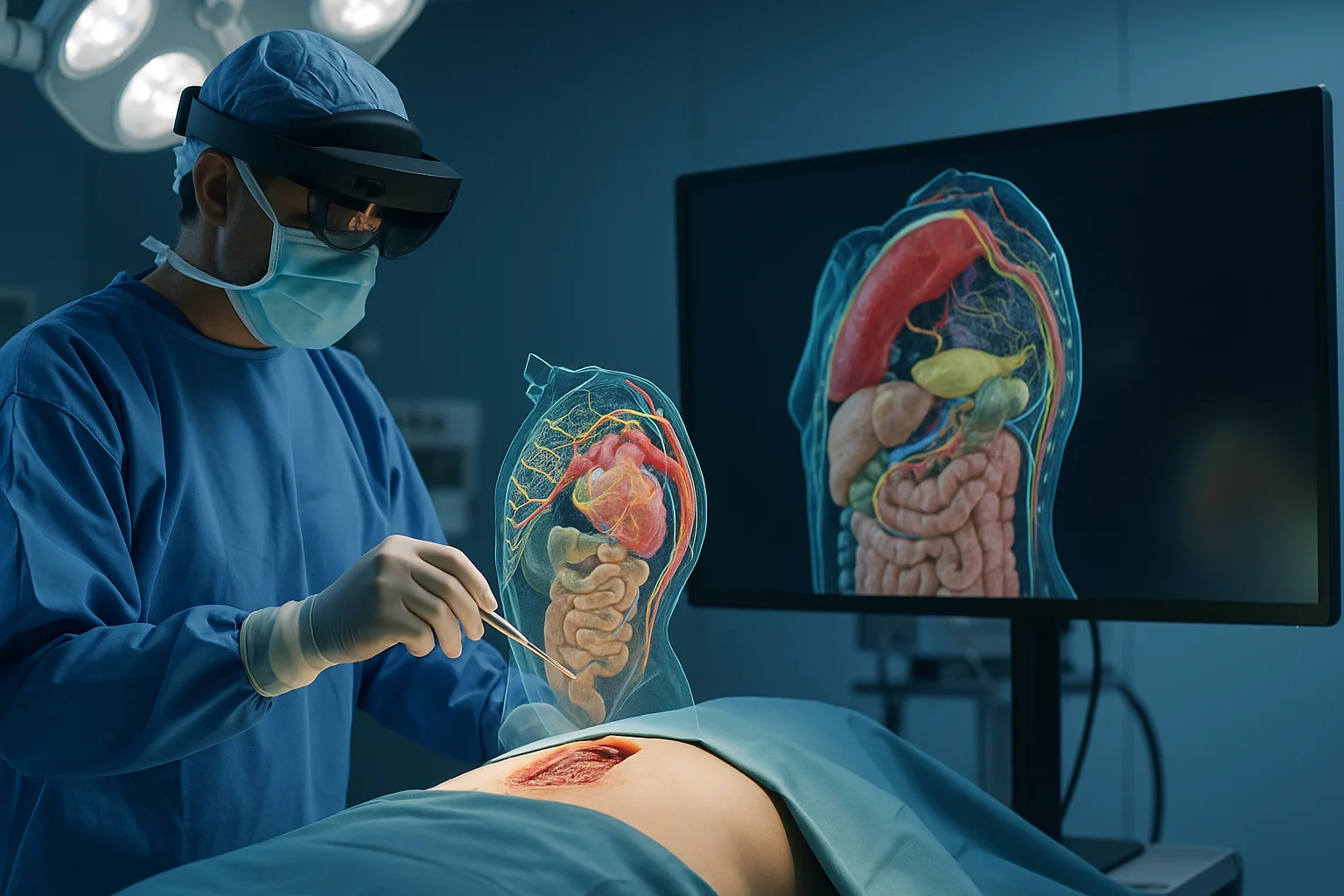
Augmented Reality and 3D in Surgical Procedures
Augmented reality and 3D modeling are transforming how surgical procedures are planned, performed and taught, giving surgeons a digital window into the patient's anatomy.
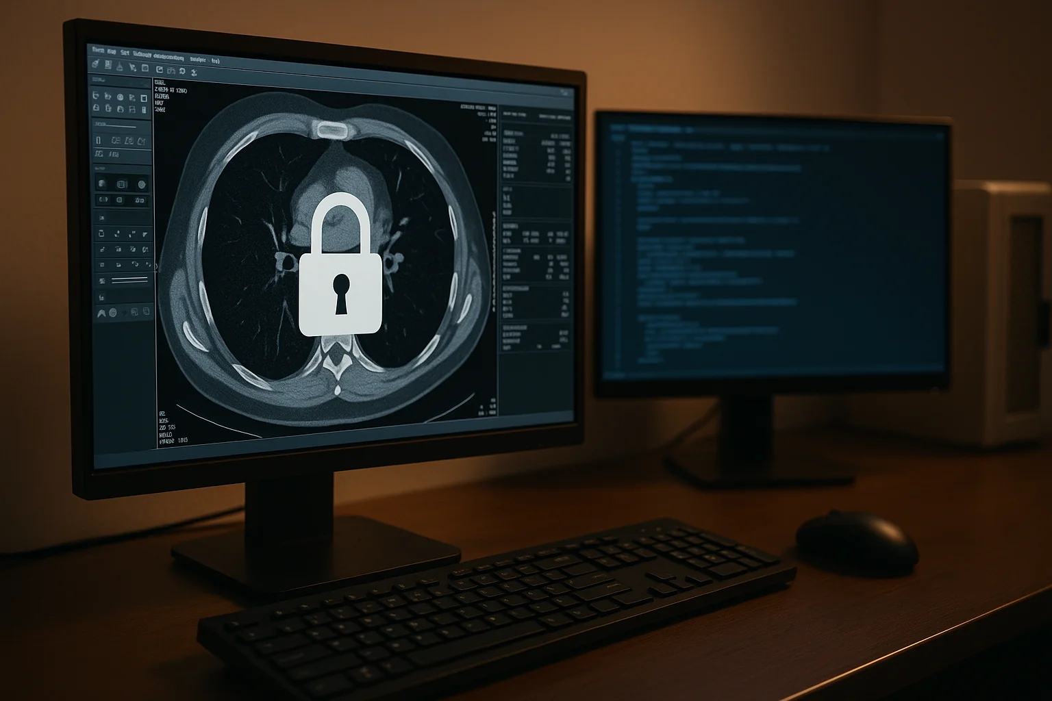
Data Security in Digital Radiology
Digital radiology handles sensitive images, predictive models and valuable clinical data. Protecting this information is essential to ensure safe diagnoses, patient trust and operational continuity.
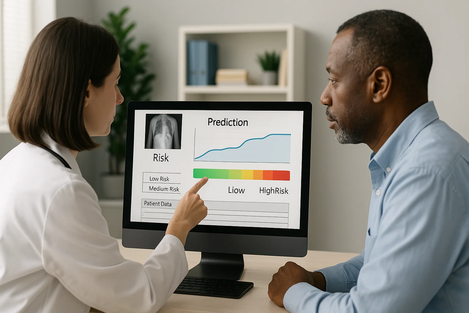
Image-Based Predictions: Medicine That Stays One Step Ahead
Thanks to artificial intelligence, medical images no longer just show what is happening, but what could happen. Predictive medicine turns every study into a tool to anticipate risks and save lives ahead of time.