
How Deep Learning Improves MRI and CT Scans
How deep learning improves MRI and CT scans
In the previous article, “Report automation: the new era of the digital radiologist” , we explored how artificial intelligence is helping radiologists produce faster, more accurate and more consistent reports. But the advances are not limited to interpretation or documentation: today AI also acts directly on image quality. The star of this evolution is deep learning.
When machines learn to improve what they see
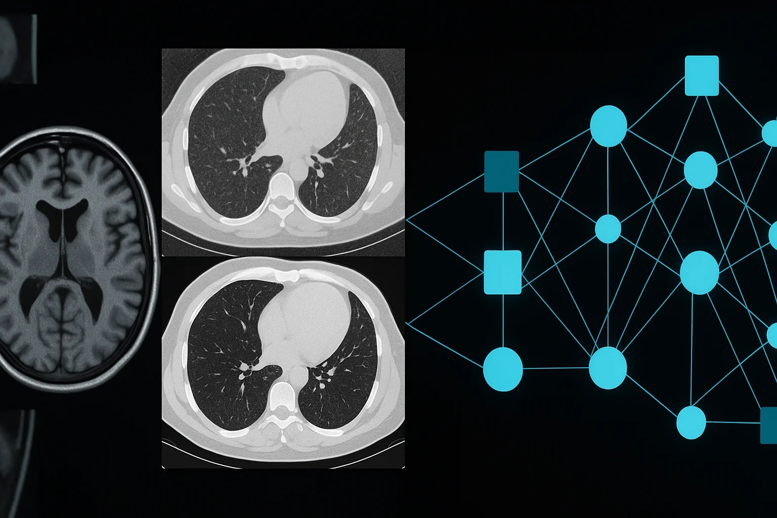
Deep learning is an advanced branch of machine learning based on deep neural networks. These networks can learn complex patterns from millions of medical images. The remarkable thing is that they do not just interpret: they also reconstruct and optimize magnetic resonance imaging (MRI) and computed tomography (CT) studies.
Put simply, deep learning works like a “digital radiologist” that cleans, clarifies and enhances the image before a human sees it. The result is unprecedented diagnostic accuracy.
Less noise, more detail
One of the biggest challenges in CT and MRI is the noise that appears in images due to patient movement, long acquisition times or low radiation doses. Neural networks learn to identify that noise and remove it without erasing important details.
This enables:
- Sharper images even with low radiation doses.
- Significantly shorter scan times in MRI.
- Fewer repeat studies due to poor quality or movement.
Deep learning does not just improve appearance: it improves clinical reliability.
Accelerated image reconstruction
In MRI, deep learning has made it possible to cut scan times that used to last 20 minutes to just 3 or 4, without losing definition. This is achieved through accelerated reconstruction techniques: the machine acquires less data, and the algorithm reconstructs the final image with equal or higher quality.
This innovation:
- Reduces waiting lists.
- Increases patient comfort.
- Optimizes the use of expensive equipment.
Technology is democratizing access to advanced studies.
Better diagnoses, less radiation
In CT, the use of AI makes it possible to reduce the radiation dose by up to 40–60% while maintaining superior quality. Algorithms reconstruct what is missing, fill in incomplete data and smooth out artifacts without compromising accuracy.
This is especially valuable in:
- Pediatric patients.
- Repeated studies for cancer follow-up.
- Screening programs such as lung cancer screening.
AI does not just improve the image: it makes it safer.
Deep learning inside PACS and RIS
As we saw in previous articles, PACS and RIS systems already integrate artificial intelligence tools. Deep learning is being built directly into these systems, so that:
- Images are automatically optimized when they are uploaded.
- Defective studies are identified and selective repeats are recommended.
- Image series are intelligently tagged according to suspicious findings.
It is a faster, more transparent workflow with fewer human errors.
Explainable AI: essential in image enhancement
As discussed in explainable AI, transparency remains essential. When an algorithm alters or enhances a medical image, the radiologist must know how and why it did so. That is why modern systems include:
- Heat maps of the reconstruction process.
- Confidence indicators for the algorithm.
- Image versioning (original vs. enhanced).
Enhancement must never compromise accuracy. AI must be an ally, not an opaque filter.
The future: intelligent images in real time
What looks like innovation today will very soon be standard. The MRI and CT scanners of the future will include native AI capable of:
- Correcting movement in real time.
- Generating ultra-HD images from fast sequences.
- Customizing parameters according to the patient's phenotype.
- Automatically alerting about critical findings.
Medical imaging will be faster, clearer and more predictive than ever.
Conclusion
Deep learning is transforming the way MRI and CT scans are generated and analyzed. It does not just improve visual quality: it improves the quality of diagnosis. It makes studies faster, safer and more accessible. And it paves the way for a future in which radiology will be a perfect blend of digital speed and human judgment.
← Previous: Report automation: the new era of the digital radiologist
Next: Fusing imaging + clinical data: integrated diagnosis →
Related Articles
Keep exploring similar content.
Augmented Reality and 3D in Surgical Procedures
Augmented reality and 3D modeling are transforming how surgical procedures are planned, performed and taught, giving surgeons a digital window into the patient's anatomy.
Data Security in Digital Radiology
Digital radiology handles sensitive images, predictive models and valuable clinical data. Protecting this information is essential to ensure safe diagnoses, patient trust and operational continuity.
Image-Based Predictions: Medicine That Stays One Step Ahead
Thanks to artificial intelligence, medical images no longer just show what is happening, but what could happen. Predictive medicine turns every study into a tool to anticipate risks and save lives ahead of time.